Upper Thigh Anatomy : Lower Extremities Arteries And Nerves Anatomy Branches Kenhub - The four muscles all extend the lower leg.. I grew up playing backyard football. It was great fun and certainly a lot of exercise. The posterior upper leg muscles provide your knees with mobility (extension, flexion and rotation) and strength. Anatomically, it is part of the lower limb. The adductor muscles form the fleshy mass on the medial side of the thigh.
The thigh bears much of the load of the body's weight when a person is upright. Like the adductors, the abductors are also responsible for stabilizing your knees during athletic and everyday movement. The hip muscles encompass many muscles of the hip and thigh whose main function is to act on the thigh at the hip joint and stabilize the pelvis.without them, walking would be impossible. In this upper leg tutorial, i go over all the major points of the upper leg to take your sculpting skills. Female upper thigh anatomy / how to treat adductor tendonitis the art of manliness / we look at the associated symptoms and treatment options.
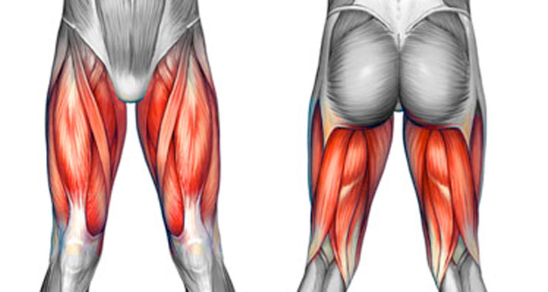
Upper Leg And Lower Leg Muscle Anatomy from www.anatomynote.com The muscles located within the posterior compartment of the thigh are the biceps femoris, semitendinosus and semimembranosus. 2, vastus medialis & intermedius muscles. It is thin and flattened, broad above, narrow and tapering below. In clinical anatomy the thigh muscles are divided into three groups: Muscles play an important role in the. They have a lot to do with how your hips move. Ebraheim's educational animated video describes muscle anatomy of the thigh. Anatomically, it is part of the lower limb.
In clinical anatomy the thigh muscles are divided into three groups:
Anatomy of the thigh : Sartorius, and the four quadriceps muscles; The adductor muscles form the fleshy mass on the medial side of the thigh. The rectus femoris is located in the center of the thigh, while the vastus medialis is in the middle of the said body part. They work closely with your quadriceps muscles at the front of your thigh, your gluteal muscles, and your calf muscles to ensure proper movement of your leg and hip. The muscles in the upper leg power many of our movements. Iliopsoas muscle, a hip flexor muscle that attaches to the upper thigh bone. Legs are used for standing, and all forms of. The single bone in the thigh is called the femur.this bone is very thick and strong (due to the high proportion of bone tissue), and forms a ball and socket joint at the hip, and a modified hinge joint at the knee. It contains many muscles and nerves but only has one bone, the femur, which is the longest and strongest bone. The hamstring portion of the adductor magnus has a similar action to these muscles, but is located in the medial thigh. Ebraheim's educational animated video describes muscle anatomy of the thigh. Female upper thigh anatomy / how to treat adductor tendonitis the art of manliness / we look at the associated symptoms and treatment options.
Muscle relaxation anatomy 12 photos of the muscle relaxation anatomy muscle relaxation anatomy, steps of muscle relaxation anatomy, human muscles, muscle relaxation anatomy, steps of muscle relaxation anatomy. Ebraheim's educational animated video describes muscle anatomy of the thigh. For example, some muscles located in the chest also help move the shoulders. The rectus femoris is located in the center of the thigh, while the vastus medialis is in the middle of the said body part. I grew up playing backyard football.
Muscles Of The Hips And Thighs Human Anatomy And Physiology Lab Bsb 141 from cnx.org It is thin and flattened, broad above, narrow and tapering below. The anatomy of the vastus medialis the vastus medialis oblique (vmo) of the quads extends the knee. The upper leg, in particular, is comprised of bones and muscles that are susceptible to injury, particularly when excess strain is placed upon them. Related posts of muscle anatomy of upper thigh muscle relaxation anatomy. It contains many muscles and nerves but only has one bone, the femur, which is the longest and strongest bone. It arises by a thin aponeurosis from the anterior margins of the lower half of the symphysis pubis and the upper half of the pubic arch. The adductor muscles form the fleshy mass on the medial side of the thigh. Legs are used for standing, and all forms of.
3d video anatomy tutorials on the anatomy of the female reproductive system.
It features two bones known as the tibia, or shin bone, and the smaller fibula.depending on their location in the anatomy of the leg, its muscle groups are divided into four different regions called compartments. It's the area that runs from the hip to the knee in each leg. January 30, 2021 a person with defined leg muscles. The muscles located within the posterior compartment of the thigh are the biceps femoris, semitendinosus and semimembranosus. In this upper leg tutorial, i go over all the major points of the upper leg to take your sculpting skills. Your legs are two of your most important body parts. Iliopsoas muscle, a hip flexor muscle that attaches to the upper thigh bone. The iliacus muscle continues down through the pelvis and attaches to the small piece of bone (lesser trochanter) that is attached to your femur (upper thigh bone). It contains many muscles and nerves but only has one bone, the femur, which is the longest and strongest bone. Ebraheim's educational animated video describes muscle anatomy of the thigh. Rectus femoris muscle, one of the quadriceps muscles on the front of your thigh. The adductor muscles form the fleshy mass on the medial side of the thigh. Muscle anatomy of upper thigh, human muscles, muscle anatomy of upper thigh.
Muscles play an important role in the. Sartorius, and the four quadriceps muscles; The fibers run vertically downward, and end in a rounded tendon, which passes behind the medial condyle. Anatomy of the thigh : They have a lot to do with how your hips move.
Thigh Pain Injuries Symptoms Causes And Treatment from www.sportsinjuryclinic.net The muscles in the upper leg power many of our movements. Or vmo) is one of the four quadriceps muscles in the front of your upper thigh. 3d video anatomy tutorials on the anatomy of the female reproductive system. The iliacus muscle continues down through the pelvis and attaches to the small piece of bone (lesser trochanter) that is attached to your femur (upper thigh bone). There are five muscles in the anterior thigh compartment: Spine (vertebral column) upper leg (thigh) other muscles beyond the back also help move the head, shoulders, arms, and legs. Human anatomy atlas, anatomy games and cards. They have a lot to do with how your hips move.
The muscles located within the posterior compartment of the thigh are the biceps femoris, semitendinosus and semimembranosus.
It is thin and flattened, broad above, narrow and tapering below. •medial thigh muscles•adductor longus muscle•adductor magnus muscle•adductor. Legs give us the freedom to run, walk, jump, climb, and negotiate the world around us. It contains many muscles and nerves but only has one bone, the femur, which is the longest and strongest bone. Muscle anatomy of upper thigh, human muscles, muscle anatomy of upper thigh. Muscle relaxation anatomy 12 photos of the muscle relaxation anatomy muscle relaxation anatomy, steps of muscle relaxation anatomy, human muscles, muscle relaxation anatomy, steps of muscle relaxation anatomy. The hip muscles encompass many muscles of the hip and thigh whose main function is to act on the thigh at the hip joint and stabilize the pelvis.without them, walking would be impossible. The thigh bears much of the load of the body's weight when a person is upright. Anatomically, it is part of the lower limb. Meanwhile, the vastus lateralis is on the side of the thigh, while the vastus intermedius is hidden below the rectus femoris(5). In clinical anatomy the thigh muscles are divided into three groups: On the anterior side, the most prominent of the muscles are the sartorius muscle and the four muscles that make up quadriceps muscle group (the quads.) Sartorius, and the four quadriceps muscles;


0 Comments:
Posting Komentar